

Hidden in the Folds: The Hidden Epidemic of Intertriginous Wounds and How to Heal Them
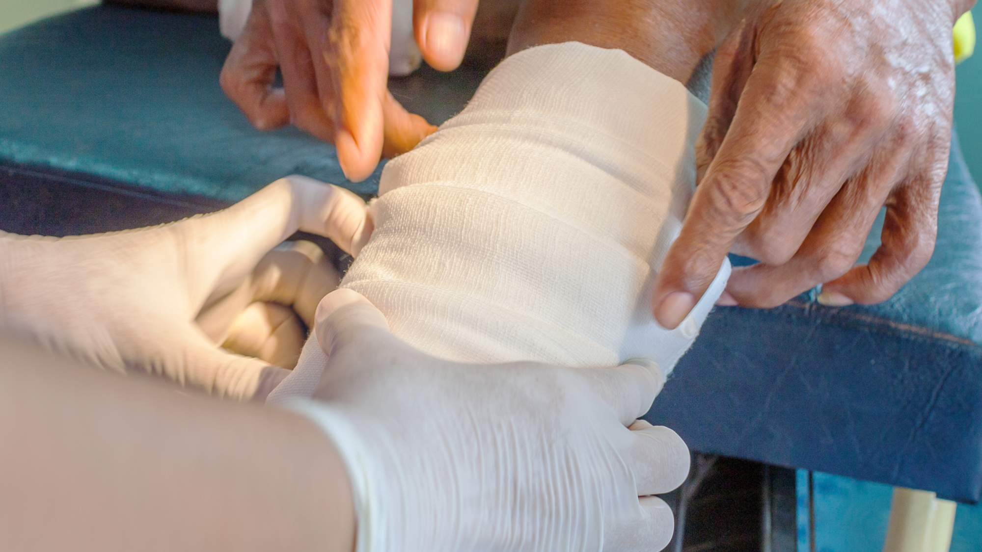
When discussing advanced wound care, public attention usually gravitates toward visible injuries like diabetic foot ulcers or surgical incisions. Yet, there is a silent, agonizing category of wounds that patients often hide out of embarrassment: intertriginous wounds.
These are wounds that develop deep within the natural folds of the body, under the breasts (inframammary), within abdominal folds (panniculus), in the groin (inguinal), or between the buttocks. Because these areas are hidden from plain sight, early stage damage is easily missed, allowing simple skin irritation to rapidly degenerate into painful, weeping, and complex chronic wounds (Sibbald et al., 2021).
At Vertex Wound Specialists, we believe that no wound is too hidden to deserve expert, empathetic, and advanced restorative care.
The Anatomy of Conflict: Friction, Moisture, and Microbes
To understand why intertriginous wounds are so difficult to manage, we have to look at the unique microenvironment of a skin fold. This condition begins as Intertriginous Dermatitis (ITD), a subset of Moisture-Associated Skin Damage (MASD) (Kottner et al., 2020).
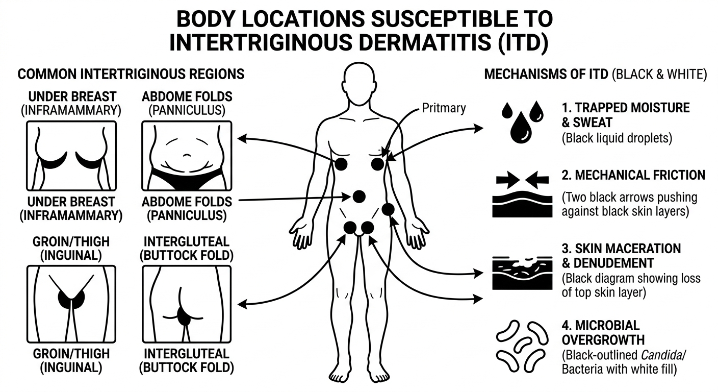
Common areas susceptible to intertriginous wound development
When two opposing skin surfaces trap sweat and body oils, it creates a cascade of physiological damage:
Skin Maceration: Prolonged exposure to trapped moisture overhydrates the stratum corneum (the outermost protective layer of skin). Think of how your fingers wrinkle after a long bath; macerated skin loses its cellular integrity and becomes weak, soft, and easily torn (Beeckman et al., 2020).
Mechanical Friction: As the body moves, these softened skin layers rub against one another. This constant friction causes denudement, literally stripping away the top layers of skin.
The Microbial Shift: The human skin surface is naturally acidic, which keeps harmful microbes at bay. Trapped sweat degrades this acid mantle, shifting the pH from acidic to neutral or alkaline. This warm, moist, alkaline environment becomes a perfect incubator for opportunistic pathogens, most notably Candida albicans (yeast) and bacteria like Staphylococcus aureus (Metin et al., 2018).
Once the skin tears open under these conditions, a standard band-aid won't cut it. The wound sits in a constant state of shearing force and moisture overload, completely stalling the body's natural cellular healing timeline.
When a Rash Becomes a Complex Wound
Many patients attempt to treat these issues at home using cornstarch, heavy cosmetic lotions, or over-the-counter anti-itch creams. Unfortunately, these steps often make the problem worse. Cornstarch can clump and create further friction, while thick ointments trap more moisture, accelerating tissue breakdown (Sibbald et al., 2021).
Without specialized intervention, what started as a simple chafed rash can turn into a deep fissure, an ulceration, or a systemic bacterial skin infection (cellulitis).
The Vertex Protocol: Advanced Management for Skin Fold Wounds
At Vertex Wound Specialists, our team utilizes an objective, multi-step therapeutic framework that goes far beyond surface-level topicals to address the root causes of intertriginous breakdown.
Moisture Mgmt. & Wicking ➡️Microbial Eradication ➡️Tissue Regeneration & Shearing Reduction
Moisture Mgmt. & Wicking ➡️Microbial Eradication ➡️Tissue Regeneration & Shearing Reduction
1. Specialized Hydrophobic & Polymer Barriers
Instead of thick creams that trap fluid, we utilize advanced liquid skin protectants. These specialized formulations create a transparent, breathable, and long-lasting polymeric barrier over denuded tissue. It shields the raw nerve endings and exposed dermis from friction and moisture while allowing the wound to "breathe" and heal (Beeckman et al., 2020).
2. Moisture-Wicking Textiles with Antimicrobial Silver
Managing the microclimate inside a skin fold is essential. We utilize highly specialized, breathable woven fabrics embedded with silver ions. These textiles actively wick moisture away from the wound bed via capillary action, keeping the area dry while the silver continuously releases ions to kill off yeast and bacterial bio-burden (Dissemond et al., 2020).
3. Targeted Negative Pressure Wound Therapy (NPWT)
For deep, high-exudate (heavily weeping) intertriginous wounds, traditional dressings will saturate and fail within hours. In these advanced cases, we customize Negative Pressure Wound Therapy using specialized, flexible foam contours. This continuous, gentle vacuum removes excess fluid, pulls the wound edges together, and stimulates localized blood flow to rapidly build healthy new tissue (Zaveri et al., 2024).
Healing with Dignity and Expertise
Intertriginous wounds can be incredibly painful, limiting your mobility, impacting your self-esteem, and exposing you to serious infectious risks. You do not have to suffer in silence or rely on trial-and-error home remedies that fail to provide lasting relief.
Our specialized clinicians provide a discreet, comfortable, and highly advanced therapeutic environment tailored specifically to your body's unique structural and healing needs.
Don't let a hidden wound hold you back. Contact Vertex Wound Specialists today to schedule your private consultation.
References
Beeckman, D., Campbell, J., Le Blanc, K., Campbell, K., Dunk, A., El Feky, A., ... & Woo, K. (2020). International Best Practice Principles: Best practice principles for addressing moisture-associated skin damage (MASD). Wounds International.
Dissemond, J., Assadian, O., Gerber, V., Kingsley, A., Kramer, A., Leaper, D. J., ... & Schultz, G. (2020). Classification of wound dressings conformed to clinical requirements for the treatment of chronic wounds with silver. Journal of Wound Care, 29(10), 548-557. https://doi.org/10.12968/jowc.2020.29.10.548
Kottner, J., Beeckman, D., Vogt, A., & Blume-Peytavi, U. (2020). Moisture-associated skin damage: A review of its definition, etiology, and management. Journal of Wound, Ostomy and Continence Nursing, 47(6), 551-558. https://doi.org/10.1097/WON.0000000000000707
Metin, N., Dilek, N., & Demir, S. (2018). Etiological factors and clinical features in patients with intertrigo. Journal of Dermatology & Dermatologic Surgery, 22(1), 15-20. https://doi.org/10.1016/j.jdds.2017.09.001
Sibbald, R. G., Ayello, E. A., Alavi, A., Ostrow, B., & Lowe, J. (2021). Intertriginous dermatitis (intertrigo): A skin fold puzzle. Advances in Skin & Wound Care, 34(11), 572-581. https://doi.org/10.1097/01.ASW.0000790516.36872.23
Zaveri, T., Karki, S., & Shrestha, S. (2024). Negative pressure wound therapy in complex intertriginous wounds: A clinical evaluation of efficacy and patient outcomes. Journal of Wound and Regenerative Medicine, 12(2), 88-94.